Female Pelvis Anatomy Muscles / Artstation Female Pelvic Floor Anatomy Aimee Hutchinson - There are direct, transverse, oblique right and left sections of the pelvis.
Female Pelvis Anatomy Muscles / Artstation Female Pelvic Floor Anatomy Aimee Hutchinson - There are direct, transverse, oblique right and left sections of the pelvis.. These muscles origin in continuity from the body of the pubis, along a tendinous arch over the obturator internus fascia, and the ischial spine. Sagittal plane through the female pelvis. Whether it's to pass that big test, qualify for that big promotion or even master that cooking technique; Anatomy pelvis muscles pubococcygeus, puborectalis and iliococcygeus., pelvis nerve, the spinal nerves that arise from vertebral column through the sacrum., pelvic floor musculature male & female pelvis anatomy. This method, the subject of her companion volumes anatomy of movement and anatomy of movement:
Pelvic girdle and floor female pelvis and reproductive organs male pelvis and reproductive organs urinary bladder and urethra perineum the muscles are attached along the inner walls of the true pelvis to a condensed area of the obturator fascia known as the tendinous arch of levator ani muscle. This anatomy model of a female pelvis represents detailed information about the topography of bones, ligaments, pelvic floor muscles and female pelvic organs. Sartorius muscle pelvis anatomy adipose tissue radiology female. Boundaries of the pelvic outlet (anterior, lateral and posteri… male or female: Female pelvis pelvic floor muscle model uterus ovary muscle teaching resources educational supplies removable.
2 Female Pelvic Anatomy Warda Part 2 from image.slidesharecdn.com Female reproductive i and ii. This anatomy model of a female pelvis represents detailed information about the topography of bones, ligaments, pelvic floor muscles and female pelvic organs. What are the skeletal differences between these two pelves? Exercises, has been enthusiastically received in workshops that she presented for many years in france. Anatomy of the female pelvis : The muscles of the pelvis form its floor. Pelvic girdle and floor female pelvis and reproductive organs male pelvis and reproductive organs urinary bladder and urethra perineum the muscles are attached along the inner walls of the true pelvis to a condensed area of the obturator fascia known as the tendinous arch of levator ani muscle. Dummies helps everyone be more knowledgeable and confident in applying what they know.
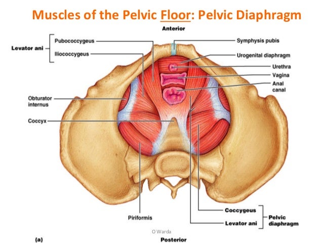
There are many muscles that form the pelvic floor, including puborectalis, pubococcygeus, iliococcygeus and coccygeus.
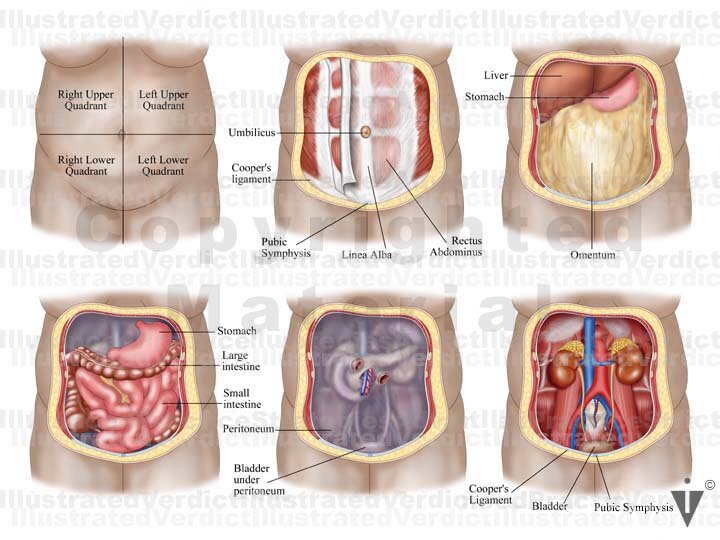
Bones of the pelvis | pelvic anatomy. Female perineal model vascular nerve pelvic floor muscle anatomical model. Study flashcards on normal female pelvis anatomy at cram.com. ƒ organs and structures of the female pelvis. Has been added to your cart. They produce and contain many eggs that mature here. Nowadays obstetric suitability of the female pelvis is assessed by ultrasound. ƒ pelvic floor dysfunction is common and. Female reproductive i and ii. The dimensions of the head of the fetus and of the birth canal are accurately measured and compared, and the feasibility of labor can be predicted. This landmark in females, the pelvis also houses the uterus, fallopian tubes, and ovaries. The best way to memorize and retain information is by spaced repetition. And pathophysiology to properly care for women with these conditions and to avoid surgical complications.
The greater or false pelvis (pelvis major).—the greater pelvis is the expanded portion of the cavity situated above and in front of the pelvic brim. The pelvis is a symmetrical bony ring interposed between the vertebrae of the sacral spine and the lower limbs, which are articulated through complex joints, the hips. Sartorius muscle pelvis anatomy adipose tissue radiology female. The pelvic floor muscles are the layer that supports the pelvic organs and spans the bottom of the pelvis. Female perineal model vascular nerve pelvic floor muscle anatomical model.
Female Pelvis Anatomical Model With Ligaments Sagittal Midsection Through The Pelvic Floor Muscles 4 Parts Genital And Pelvis Anatomical Models Anatomical Models And Plates Physiotherapy Equipment Fisaude Store from www.fisaude.eu There are many muscles that form the pelvic floor, including puborectalis, pubococcygeus, iliococcygeus and coccygeus. Mccarthy s, tauber c, gore j. In front it is incomplete, presenting a wide interval between the anterior borders of the ilia. This landmark in females, the pelvis also houses the uterus, fallopian tubes, and ovaries. Knowledge of anatomy unique to females is essential for all clinicians, especially those in the field of obstetrics and gynecology. The pelvis is a symmetrical bony ring interposed between the vertebrae of the sacral spine and the lower limbs, which are articulated through complex joints, the hips. The true pelvis, or lesser pelvis, lies below the pelvic brim (figure 1). Thus, in the standing position, the bony pelvis is ori
This section of the website will explain large and minute details of mri sagittal cross sectional anatomy of female pelvis (uterus and ovaries ).
If you're curious to know. Female pelvis pelvic floor muscle model uterus ovary muscle teaching resources educational supplies removable. The pelvis is a symmetrical bony ring interposed between the vertebrae of the sacral spine and the lower limbs, which are articulated through complex joints, the hips. This anatomy section promotes the use of the terminologia anatomica, the international standard of anatomical nomenclature. What are the skeletal differences between these two pelves? Anatomical drawing of the female pelvis. These muscles origin in continuity from the body of the pubis, along a tendinous arch over the obturator internus fascia, and the ischial spine. Pubococcygeus, puborectalis, iliococcygeus anatomy of the pelvic floor. The greater or false pelvis (pelvis major).—the greater pelvis is the expanded portion of the cavity situated above and in front of the pelvic brim. Sagittal plane through the female pelvis. The dimensions of the head of the fetus and of the birth canal are accurately measured and compared, and the feasibility of labor can be predicted. Boundaries of the pelvic outlet (anterior, lateral and posteri… male or female: Pelvic girdle and floor female pelvis and reproductive organs male pelvis and reproductive organs urinary bladder and urethra perineum the muscles are attached along the inner walls of the true pelvis to a condensed area of the obturator fascia known as the tendinous arch of levator ani muscle.
It is bounded on either side by the ilium; Muscles of the true pelvis. Nowadays obstetric suitability of the female pelvis is assessed by ultrasound. The best way to memorize and retain information is by spaced repetition. Endopelvic fascia attachments of the pelvic floor.
Stock Female Pelvis Normal Anatomy Illustrated Verdict from images.squarespace-cdn.com Floor muscles female pelvis anatomy model. Males and females differ significantly in the anatomy of the pelvis: Mccarthy s, tauber c, gore j. There are many muscles that form the pelvic floor, including puborectalis, pubococcygeus, iliococcygeus and coccygeus. The female bony pelvis is divided into: The dimensions of the head of the fetus and of the birth canal are accurately measured and compared, and the feasibility of labor can be predicted. These muscles origin in continuity from the body of the pubis, along a tendinous arch over the obturator internus fascia, and the ischial spine. The greater or false pelvis (pelvis major).—the greater pelvis is the expanded portion of the cavity situated above and in front of the pelvic brim.
Above the pelvic brim and has no obstetric importance.
Muscles of the true pelvis. It bisects the true conjugate and is slightly shorter than the anatomical transverse diameter. Dummies has always stood for taking on complex concepts and making them easy to understand. Mccarthy s, tauber c, gore j. If you're curious to know. They produce and contain many eggs that mature here. Magn reson imaging clin n am. In front it is incomplete, presenting a wide interval between the anterior borders of the ilia. What are the skeletal differences between these two pelves? Bones of the pelvis | pelvic anatomy. Female perineal model vascular nerve pelvic floor muscle anatomical model. Differences between the male pelvis and the female pelvis. Anatomy pelvis muscles pubococcygeus, puborectalis and iliococcygeus., pelvis nerve, the spinal nerves that arise from vertebral column through the sacrum., pelvic floor musculature male & female pelvis anatomy.
Female pelvis is the locationovaries, which are located on the sides of the uterus anatomy muscles pelvis. Sartorius muscle pelvis anatomy adipose tissue radiology female.


0 Komentar